
Kalvis Pastars, M.D.
Board-certified plastic surgeon, microsurgeon, hand surgeon

Skin cancer remains the most common form of cancer worldwide. Although it is more common in people over the age of 50, the incidence among younger people (up to age 30) has also been increasing recently.
The at-risk group includes people with fair skin (phototypes I and II) and light hair, people with freckles, individuals with an increased number of moles, people who spend a lot of time in the sun, and frequent tanning bed users. The main carcinogenic factor is ultraviolet radiation—these tumors mostly (>85% of cases) develop on areas of the body exposed to the sun.
Skin cancer is divided into two main groups: non-melanoma tumors (basal cell carcinoma and squamous cell carcinoma) and melanoma, which develops from pigment-producing cells called melanocytes.
Basal cell carcinoma comes in various forms with differing degrees of aggressiveness. Nodular and superficial forms are low-risk, while infiltrating forms are the most aggressive and carry a high risk of recurrence. Basal cell carcinoma very rarely metastasizes. Basal cell carcinoma is initially painless, so regular self-examination is important. Persistent redness, itching, peeling skin, a new or changing mole, or a non-healing wound or ulcer (>2 weeks) may indicate a malignant process. Basal cell carcinomas are classified as nodular, ulcerated, superficial, infiltrating, and pigmented.
In most cases, squamous cell carcinoma develops from precancerous conditions—actinic (solar) keratosis or Bowen’s disease. There is a risk of metastasis, which is increased by tumor size (especially >2 cm) and location in high-risk anatomical areas. The diagnosis is confirmed by cytological or histological analysis; regional lymph nodes must also be examined.
Melanoma is a malignant and serious skin condition that develops from melanocytes. If detected early, it can be cured. The lesion is evaluated based on its shape, borders, color, size (>6 mm), surface, and texture, as well as the “ugly duckling” sign. Types: in situ, superficial (~70%), nodular, lentigo maligna, acral lentiginous, and subungual melanoma. The only accurate diagnostic method is a biopsy followed by histological examination.
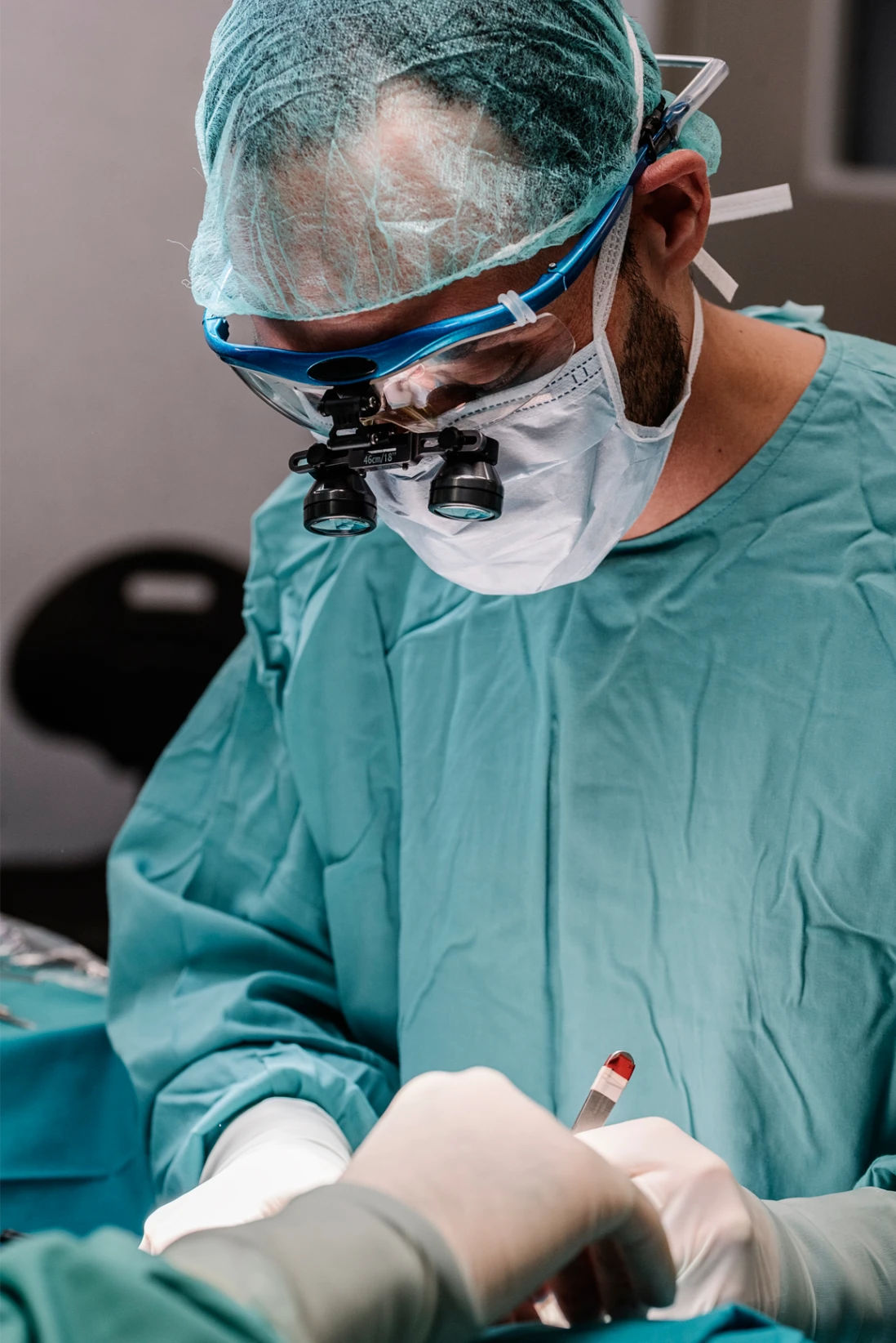
The primary treatment method is surgical excision of the lesion within the margins of healthy tissue—the “gold standard,” which, with a margin of >4 mm, ensures a cure in up to 95% cases. Additional treatments for melanoma include targeted therapy, immunotherapy, and radiation therapy. At the Latvian Microsurgery Center, it is possible to perform microsurgical excision of tumors under optical magnification with examination of the resection margins, as well as reconstructive/plastic surgery.

+
+
+